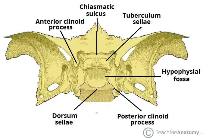
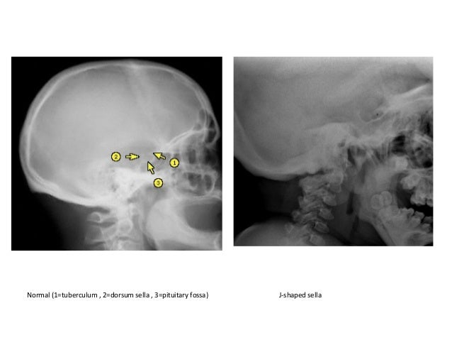
Floor View Skull Dorsum Sellae

Specialised Projections Of The Skull Radiology Key
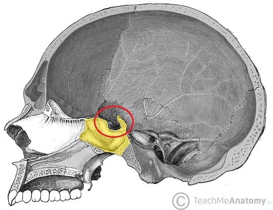
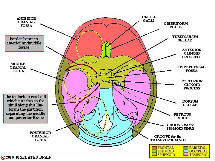
Middle Cranial Fossa Boundaries Contents Teachmeanatomy

Landmarks For Cephalometric Analysis S Sella Center Of Sella Download Scientific Diagram
Anatomy Of The Skull Base And Related Structures Elements Of Surgical Anatomy Neupsy Key

Skull X Ray Lateral View Note Enlargement Of Pituitary Fossa Loss Download Scientific Diagram
Pixelated Brain Module 1 Section 1 The Skull
In a properly positioned caldwell projection the ir is perpendicular to the orbitomeatal line oml and the x rays pass at an angle of 15 degrees from behind the head and exit at the nasion.
Floor view skull dorsum sellae.

The Axial Skeleton Flashcards Quizlet
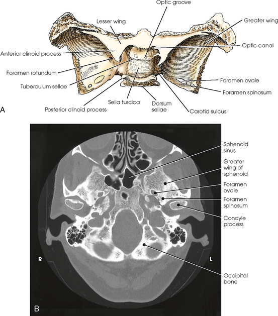
Sphenoid Bone Location Structure Function Teachmeanatomy
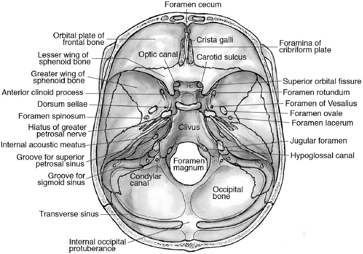
Skull Foramina Fissures And Contents Kenhub

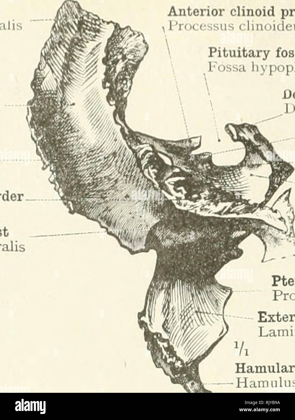
Sphenoid Bone
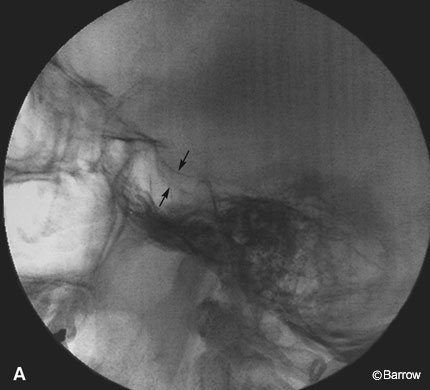
Double Sellar Floor Radiographic Sign For A Pituitary Adenoma Barrow

Procedures 3 Skull Anatomy Flashcards Quizlet
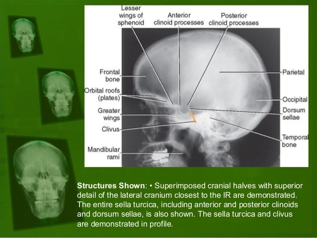
Positioning And Radiographic Anatomy Of The Skull

Imaging Of Skull Base Lesions Sciencedirect

Anterior Cranial Fossa Nasal Cavity And Paranasal Sinuses Radiology Key
Skull Radiology Key
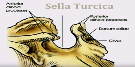
Sella Turcica Assignment Point
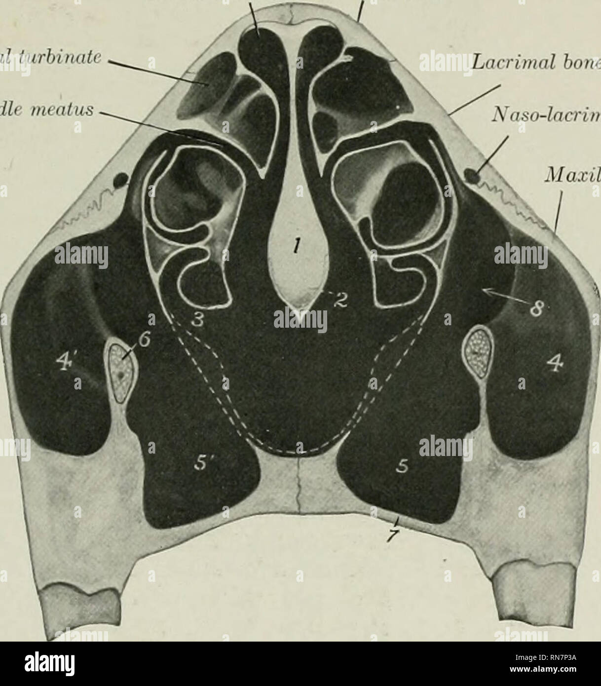
The Anatomy Of The Domestic Animals Veterinary Anatomy Skull Of The Ox As A Whole 143 Than The Rest Of The Floor The Ethmoidal Fossffi Are Smaller And The Hypophyseal Fossa

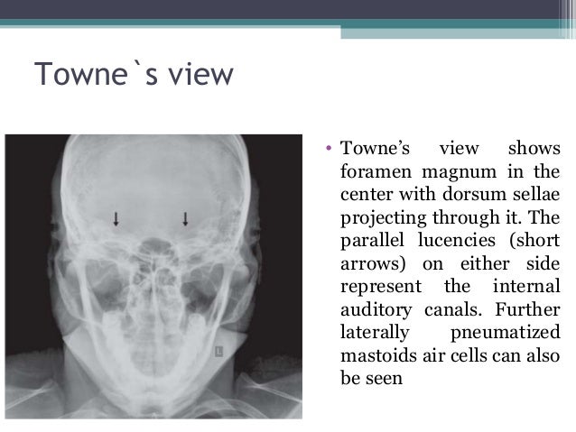
Fingerstocspine Ppt Ap Axial Skull Dorsum Sellae Projected In Foramen Magnum Entire Skull Visualized No Rotation Or Tilt Petrous Ridges Symmetric Course Hero

Osteology Of Head Neck
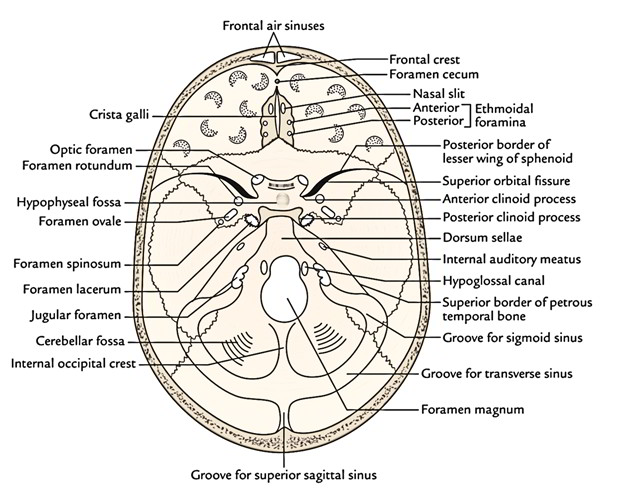
Easy Notes On Cranial Cavity Learn In Just 4 Minutes Earth S Lab
Skull X Ray Plain Evaluations
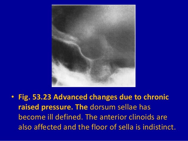
53 David Sutton Pictures The Skull

Intraoperative Photographs Of The Floor Of The Third Ventricle In Case Download Scientific Diagram
Https Encrypted Tbn0 Gstatic Com Images Q Tbn 3aand9gcsdpcg9veimnadcpwli87idy8t7twt44anlaworyjy Usqp Cau
Diagnostic Imaging Of The Pituitary Gland

Sella And Skull Base Flashcards Quizlet
Gale Academic Onefile Document Endoscopic Endonasal Transpituitary Gland Approach For Resection Of Dorsum Sellae Meningioma Technical Case Report

Ancient Skull From A 40 Year Old Female With Osteoporosis Download Scientific Diagram
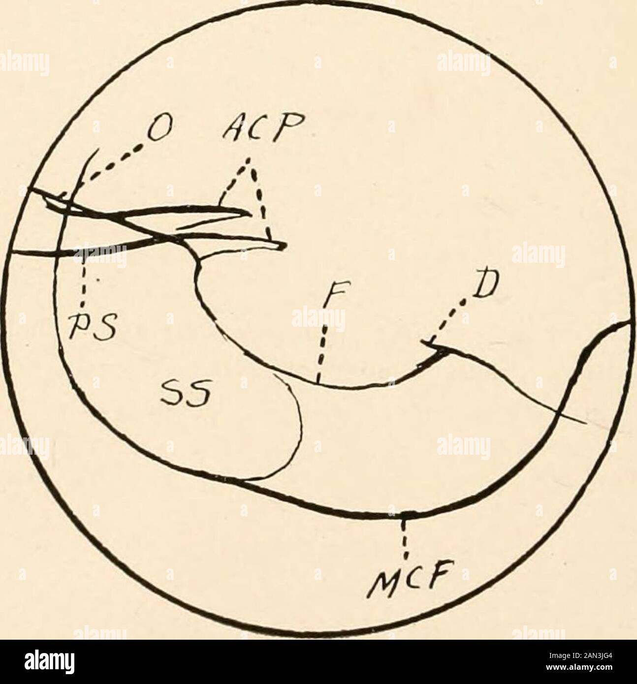
Roentgen Diagnosis Of Diseases Of The Head Fig 72 Roentgenogram Of Case 3 Page 193 The Dorsum Sell E Is Almostentirely Gone Fig 73 Sketch Of Fig 72 O Roof Of Orbit Acp Anterior
Source : pinterest.com
